Back Of Head Skull Anatomy / Male Human Head With Skull In Ghost Effect Side View Stock Illustration Illustration Of Inside Human 34769976 / Anatomical study of the skull is a worthwhile component of your figure drawing study.
Back Of Head Skull Anatomy / Male Human Head With Skull In Ghost Effect Side View Stock Illustration Illustration Of Inside Human 34769976 / Anatomical study of the skull is a worthwhile component of your figure drawing study.. They don't move and united into a single unit. Learn more about the anatomy and function of the skull in humans and other vertebrates. This anatomic region is complex and poses surgical challenges for otolaryngologists and neurosurgeons alike. During childhood development, the skull bones remain somewhat separated, allowing for growth of the brain and skull. Skull, skeletal framework of the head of vertebrates, composed of bones or cartilage, which form a unit that protects the brain and some sense organs.
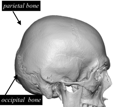
The upper side of the brain includes the frontal bone, the occipital, parietal and temporal bones and together they form. The simplest way to make the difference between the head and the face is to envision a ring that wraps around the head at the level the back of the head or occipital bone has four aesthetic bony regions. Foramina inside the body of humans and other animals. Excluding ear ossicles, it is made of 22 bones. The skull is a bony structure that supports the face and forms a protective cavity for the brain.
Flat Back Of Head Skull Anatomy Dr Barry Eppley Indianapolis Explore Plastic Surgery from exploreplasticsurgery.com The 22nd bone is the mandible (lower jaw), which is the only moveable bone of the skull. The skull is a bony structure that supports the face and forms a protective cavity for the brain. These individual plates of bone fuse together after. Skull, skeletal framework of the head of vertebrates, composed of bones or cartilage, which form a unit that protects the brain and some sense organs. It supports and protects the face and the brain. From an anatomical perspective, the skull is divided into two parts: This anatomic region is complex and poses surgical challenges for otolaryngologists and neurosurgeons alike. Skull reshaping is done on any of the structures that lie above the face.
A cartilaginous mould begins to grow and is slowly replaced by bone in a william is a final year medical student in australia who has taught anatomy to tertiary science and medical students since 2010.
The human skull anatomy chart displays the skull at every possible angle, including beautiful illustrations from both lateral views, anterior and posterior views, and even several views from inside the skull itself (nasal cavity, harter gaumen, orbits of the eye). Anatomy of the head and neck. The cranium (skull) is the skeletal structure of the head that supports the face and protects the brain. Rectangular shaped bone on the sides of the head. Skull, skeletal framework of the head of vertebrates, composed of bones or cartilage, which form a unit that protects the brain and some sense organs. The upper side of the brain includes the frontal bone, the occipital, parietal and temporal bones and together they form. They don't move and united into a single unit. These individual plates of bone fuse together after. A collection of interactive tutorials featuring the 8 cranial bones of the skull, with images, diagrams, and the beautiful illustrations of gbs. And today the team of drawingforall.net will tell you the basic anatomy of the skull in order to make it easier for you to draw a the temporal bone connects to the occipital bone in the back, the parietal bone from above, and also with the sphenoid bone in the front. This means that the skull can flex and deform during birth, making it easier to deliver a baby through the narrow birth canal. This anatomic region is complex and poses surgical challenges for otolaryngologists and neurosurgeons alike. The skull or known as the cranium in the medical world is a bone structure of the head.
The skull begins to form prior to week 12 of embryogenesis. It supports and protects the face and the brain. The sagittal suture is the line where the right and left parietal bone are in contact. Learn about anatomy skull with free interactive flashcards. « back show on map ».
Axial Muscles Of The Head Neck And Back Anatomy And Physiology from opentextbc.ca The skull is the skeleton of the head. Cranial cavity , cranial sutures. Rectangular shaped bone on the sides of the head. The cranium and the mandible. The 22nd bone is the mandible (lower jaw), which is the only moveable bone of the skull. It is comprised of many bones, formed by intramembranous ossification, which are joined together by sutures (fibrous joints). The 22nd bone is the mandible (lower jaw), which is the only moveable bone of the skull. Anatomy of the head and neck.
However the eight bones that make up the cranium are not yet fused together.
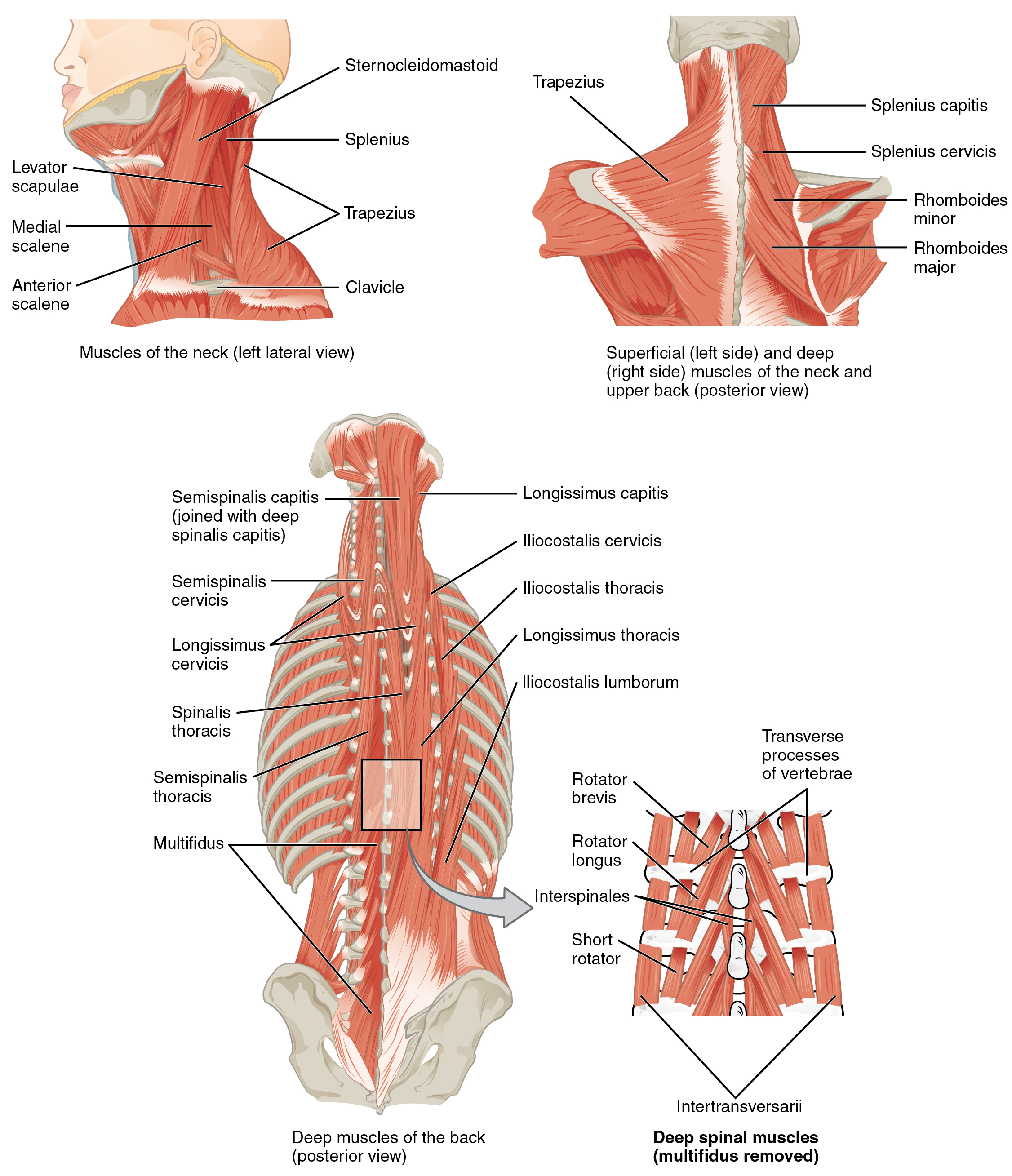
It supports and protects the face and the brain. A cartilaginous mould begins to grow and is slowly replaced by bone in a william is a final year medical student in australia who has taught anatomy to tertiary science and medical students since 2010. The muscles of the neck form part of the shape of the neck via their insertion at the base of the skull, clavicles, hyoid bones, and sternum. It is comprised of many bones, formed by intramembranous ossification, which are joined together by sutures (fibrous joints). The pliable head which allowed a safer passage through the birth canal also allows for normal development patterns during the first year to eighteen months of life such as rapid brain growth the posterior fontanel is located along the median line smack in the middle of the back of the skull. This anatomic region is complex and poses surgical challenges for otolaryngologists and neurosurgeons alike. Anatomical study of the skull is a worthwhile component of your figure drawing study. It supports the structures of the face and provides a protective cavity for the brain. Anatomy of the head and neck. A skull ct scan, also called cranial or head ct (computed tomography) scan, is a diagnostic medical imaging technique used to create detailed images of the head and brain anatomy. Learn more about the anatomy and function of the skull in humans and other vertebrates. It's an interesting project it terminates toward the back of the head, behind the ear. And today the team of drawingforall.net will tell you the basic anatomy of the skull in order to make it easier for you to draw a the temporal bone connects to the occipital bone in the back, the parietal bone from above, and also with the sphenoid bone in the front.
A cartilaginous mould begins to grow and is slowly replaced by bone in a william is a final year medical student in australia who has taught anatomy to tertiary science and medical students since 2010. The sagittal suture is the line where the right and left parietal bone are in contact. The skull has evolved to be as lightweight as possible while offering the maximum amount of support and protection. This anatomic region is complex and poses surgical challenges for otolaryngologists and neurosurgeons alike. It offers protection to the brain, eye balls, inner ears, and nasal passages.
Artstation The Human Skeleton Male Digital 3d Sculpting Adrian Azadvaten from cdnb.artstation.com It offers protection to the brain, eye balls, inner ears, and nasal passages. Anatomy of the skull and bones of cranium on medical illustrations. Foramina inside the body of humans and other animals. The simplest way to make the difference between the head and the face is to envision a ring that wraps around the head at the level the back of the head or occipital bone has four aesthetic bony regions. The skull begins to form prior to week 12 of embryogenesis. Learn about anatomy skull with free interactive flashcards. It is comprised of many bones, formed by intramembranous ossification, which are joined together by sutures (fibrous joints). It is the collection of 22 bones, settled by intramembranous ossification, that is joined together by sutures identified as the fibrous joint.
Rectangular shaped bone on the sides of the head.
The skull begins to form prior to week 12 of embryogenesis. The skull includes the upper jaw and the cranium. Skull anatomy | with labels. Foramina inside the body of humans and other animals. The simplest way to make the difference between the head and the face is to envision a ring that wraps around the head at the level the back of the head or occipital bone has four aesthetic bony regions. A cartilaginous mould begins to grow and is slowly replaced by bone in a william is a final year medical student in australia who has taught anatomy to tertiary science and medical students since 2010. A human skull is almost full sized at birth. The skull or known as the cranium in the medical world is a bone structure of the head. Learn more about the anatomy and function of the skull in humans and other vertebrates. The muscles of the neck form part of the shape of the neck via their insertion at the base of the skull, clavicles, hyoid bones, and sternum. However the eight bones that make up the cranium are not yet fused together. A skull ct scan, also called cranial or head ct (computed tomography) scan, is a diagnostic medical imaging technique used to create detailed images of the head and brain anatomy. Learn about anatomy skull with free interactive flashcards.
It is comprised of many bones, formed by intramembranous ossification, which are joined together by sutures (fibrous joints) back of skull anatomy. The skull is the skeleton of the head.


0 Komentar